INIF FAQs
Find out more about these terms, conditions and procedures:
- Neuro Intervention
- Stroke
- Cerebral Aneurysm
- Cerebral Angiograms
- Treatment of Neurovascular Diseases
- Arteriovenous Malformations (AVMs)
Neuro Intervention
What is Neuro Intervention?
Neurointervention provides a complete spectrum of minimally invasive services for the diagnosis and treatment of patients with vascular problems related to the brain and spinal cord. In the past, certain conditions that would have required open surgery such as aneurysms, carotid artery disease, vascular malformations and tumors of the head, brain, neck, and spine are now treated by Neurointervention technique.
Neurointerventional techniques such as thromboaspiration and stentrievers are also useful in the management of certain patients with acute ischemic stroke who can be treated within six (6) hours of the onset of symptoms. Neurointerventional procedures now also allow the treatment of previously untreatable or surgically difficult conditions.
Who is a Neurointerventionalist?
A neurointerventionalist is a doctor who is specially trained in neurovascular diseases and imaging. A neurointerventionalist specializes in treating conditions through minimally invasive methods.
What are the types of Neurointerventional procedures?
Following are the various types of Neurointerventional procedures:
- Acute Ischemic stroke treatment
- Cerebral aneurysms
- Treatment of TIA (Transient Ischemic Attacks) and Carotid Artery Disease
- Arteriovenous malformations (AVMs)
- Vascular malformations of head, face and neck
- AVMs and AVFistula of the Brain and Spinal cord
- Tumors of the head and neck
Stroke
What is a stroke?
Your brain gets blood mainly through two arteries in your neck (carotid arteries) and two arteries near your spine (vertebral arteries). They branch into other blood vessels that supply your brain with blood. A stroke takes place any of these blood vessels get ruptured or blocked. This cuts off or interrupts the blood and oxygen flow to the brain. Without blood, brain cells either get damaged or die. This damage can result in different effects depending on where it happens in the brain. Because of the stroke, a person may not be able to talk, see, understand and may also get paralyzed.
What are the different types of stroke?
There are two types of stroke - Hemorrhagic and Occlusive. A primary concern with both types of stroke, is to determine when the person was last known to be "normal" or without symptoms, as some treatments have time restraints.
Hemorrhagic Stroke occurs when a weakened blood vessel ruptures in the brain. Blood spills into the brain, killing tissue and cells. 15% of all strokes are hemorrhagic. During the treatment of such a stroke, it is important to:
- Determine the cause of the hemorrhage:
- Aneurysm
- AVM
- Trauma
- AVF
- Determine which vessel was the source of the hemorrhage, if possible
- Determine the best way to treat it
Occlusive/Ischemic Stroke occurs due to a blockage within a blood vessel supplying blood to the brain. This may happen because of plaque buildup in the vessels. 85% of all strokes are ischemic.
Ischemic stroke is a preventable disease in many cases. Know your risks and what you can do to reduce them, as it may result in loss of function & paralysis.The amount of damage to the brain cells is related to the level of blood flow and the length of time the blood vessel is occluded.
- The amount of tissue that has been injured but not destroyed is dependent on the quality of the secondary blood vessels that supply that area and the length of time that the primary blood vessel is occluded
- Thromboaspiration or clot retrieval is the course of treatment for occlusive stroke. The quicker the blood vessel is opened up, the fewer brain cells destroyed or injured.
- IV thrombolysis is the first line of thrombolytic therapy. It is where a drug is given intravenously that is thrombolytic or clot destroying. It must be given before 3 hours has elapsed since the person was last known to be free of symptoms and it is the only approved treatment.
- IA (Intraarterial) therapy must be given before 6 hours has elapsed since the person was last known to be symptom free. It may have a higher rate of success. It is a procedure of choice in Large Vessel Disease (ICA, MCA, Basilar arteries).
Cerebral Aneurysm
What is an Aneurysm?
An aneurysm is a bubble formed due to weakening of the blood vessel wall. The weakened area is at risk for rupture or bleeding, which can cause a brain hemorrhage. The probability of bleeding is unknown for any given individual, but statistics suggest at the most a 1% chance of bleeding each year that you live with an aneurysm - and the likelihood is ever-increasing; in 10 years, you have a 10% chance of experiencing a rupture and hemorrhage from your aneurysm. If you have more than one aneurysm, or have had bleeding from an aneurysm, your risk is much greater. 5% of the general population harbors a brain aneurysm.
What causes an Aneurysm?
Causes of aneurysm may be:
- Hemodynamic stress leading to a weak area in the wall of the artery.
- High blood pressure can cause the weakened area of the artery to bulge.
- Traumatic injury can weaken or damage the artery's wall until an aneurysm forms.
What does it look like?
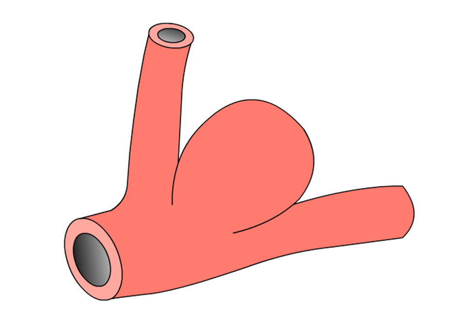
Berry Aneurysm
The most common type of aneurysm is a berry aneurysm. It is rounded like a berry and connected to the artery by a stalk or neck where the artery branches off.
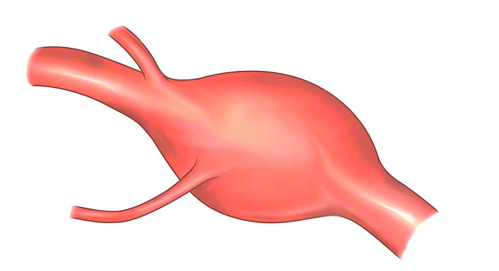
Fusiform Aneurysm
A less common type of aneurysm is a fusiform aneurysm. It is spindle-shaped. It rarely bleeds but can cause problems when it puts pressure on nearby brain tissue or nerves.
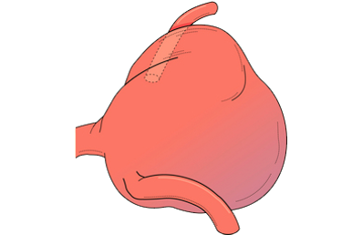
Giant Aneurysm
Another type of aneurysm is a giant aneurysm. It is like a berry aneurysm but is large, (an inch or more in diameter.) There are other less common types of aneurysms.
Cerebral Angiograms
What is a Cerebral Angiogram?
A cerebral angiogram is a test that uses dye in the blood stream and X-ray images to show abnormalities of the blood vessels in and around the brain. Some abnormal findings include narrowing, blockage from plaque or blood clots, or malformations like aneurysms. A cerebral angiogram is the most accurate test for blood vessel abnormalities and is performed before any interventional surgery is planned.
How is an Angiogram done?
A catheter tube is inserted in an artery in the groin. A special dye is injected into the blood vessels leading to your brain and X-rays are taken to show the blood vessels. The test is not painful. You will be given medicine to help relieve anxiety and to keep you comfortable while the x-rays are being taken; it takes about half an hour to one hour to finish the procedure. You will require close observation by the nurse in the hospital for two to six hours after your procedure. Your family or friend can visit during this time. You will be admitted for a day for Cerebral Angiography.
Are there any possible complications of Cerebral Angiograms?
Cerebral Angiogram is a very safe procedure that has been used since the 1930's. There is very little risk involved when performed by an experienced physician. Nevertheless, unforeseeable complications include, but are not limited to, stroke, infection or allergic reaction to dye.
Treatment of Brain Lesion: Angioplasty
What is cerebral angioplasty?
Angioplasty is a procedure that requires an angiogram to be done first. Abnormally narrow areas of the blood vessels are gently pushed open using a small, soft balloon at the end of the angiogram catheter. If needed, a stent (metal mesh cylinder) may be used to keep the expanded area open.
How is cerebral angioplasty done?
After a diagnostic angiography, any narrowing, or blockage from plaque or blood clots in the blood vessels of the brain can then be measured using X-ray images and specialized computer software. The procedure is not painful. You will be given medicine to help relieve anxiety and to keep you comfortable. In some cases, it is necessary to use general anesthesia to prevent even slight movement during the angioplasty procedure. The procedure takes two hours or more to complete, this really depends upon how tortuous, or curvy your blood vessels are. You will require close observation by the nurses in the neuro intensive care unit for 24 hours after your procedure. Standard hospitalization is for 3-4 days.
Are there any possible complications of Cerebral Angioplasty?
Uncommon (less than 2%) complications include, but are not limited to, stroke, bleeding or infection, and allergic reaction to dye.
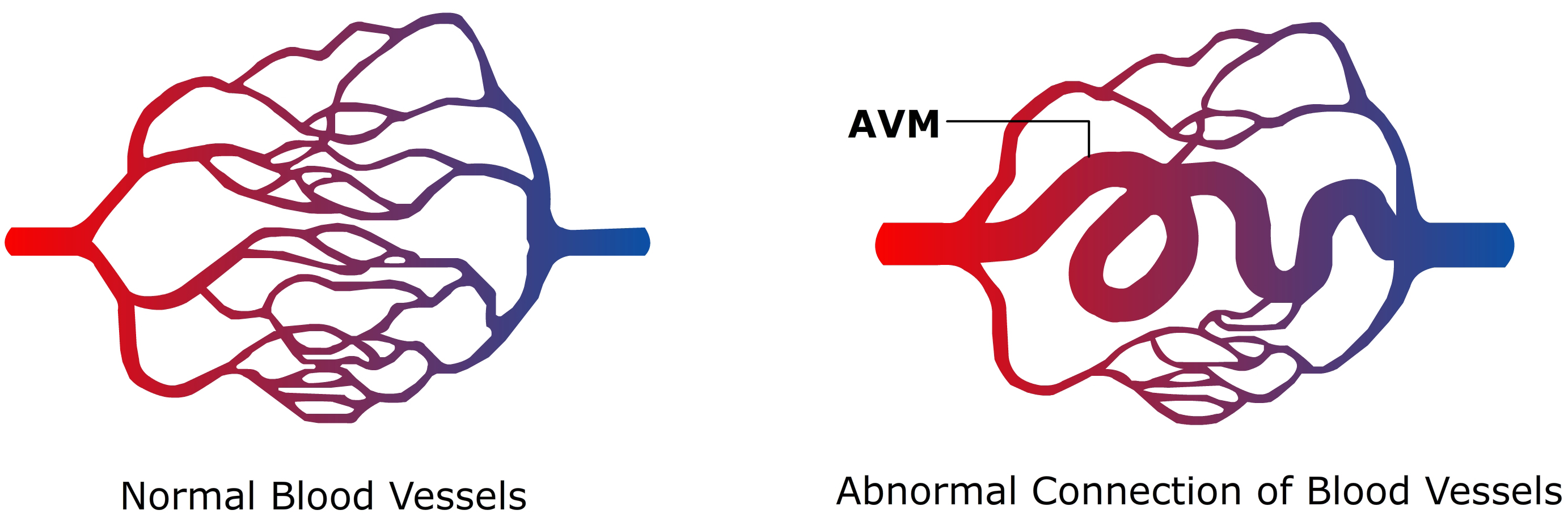
Arteriovenous Malformations (AVMs)
What are Arteriovenous Malformations (AVMs)?
Abnormal groups of tangled blood vessels known as AVMs can occur in the brain or the spine. A neurointerventionalist is able to navigate through the arteries of the brain and spinal cord to inject a fluid, which blocks the cluster nidus of the AVM.